Germination
and Anamorphs
Anamorphs in Sarcoscypha serve as valuable characters
to delimit species. Conidal size strongly varies among the species, also the guttules
inside and the nuclear numbers. Conidiophores emerging on conidiophores borne
on the ascospores is a key character for S.
austriaca and S. occidentalis.
Molliard (1904) was the
first to report conidia in Sarcoscypha. Paden (1984) erected the genus Molliardiomyces
to accommodate the anamorphic states of both Sarcoscypha and the related
genus Phillipsia, and Harrington (1990) followed this nomenclatural
method by creating further specific epithets. Thereby, the confusion about the
teleomorphs of S coccinea agg. had
the consequence that the anamorph of S. austriaca is M. coccinea while that of S.
coccinea received the name M. eucoccinea. (In my
opinion, naming of anamorphic states is not necessary after the connection to a
teleomorph has been proved by pure culture. Yet, I accept the use of form
genera to circumscribe morphologically similar anamorphic states.)
Germination
of ascospores. On current agar media
ascospores germinate readily at 15-18°C within 1-4 days (Harrington 1990: 427)
or even faster by forming usually one terminal germ tube. Multispore cultures
rapidly produce an abundant whitish mycelium.
Formation
of the anamorphic state. Possibly
all species of Sarcoscypha produce an
anamorph in pure culture. Conidia are formed on simple conidiophores. These
emerge either on the hyphae after a mycelium is produced on the agar, or
directly on the ascospores (formed on one to four loci on the ascospore wall).
The latter type is merely obtained under the absence of nutrients, either in
water agar, or when apothecia get senescent. On rich agar the ascospores always
showed normal germination, forming their conidia eventually on the mycelial
hyphae. Both types of germination occur also in the related genus Phillipsia.
Conidiogenesis is always holoblastic. Conidia produced on the ascospores are
also found inside the dead asci of senescent apothecia since ascospores need
not to be ejected in order to germinate (but they never germinate within the
living asci!).
|
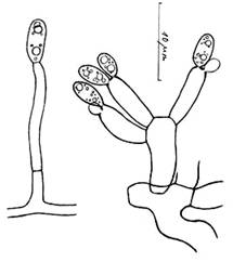
Molliardiomyces coccinea, anamorph of S.
austriaca. Conidia medium-sized, multiguttulate, ?5-9-nucleate. |
Molliardiomyces
eucoccinea, anamorph of S. coccinea. Conidia small, biguttulate, ?uninucleate. |
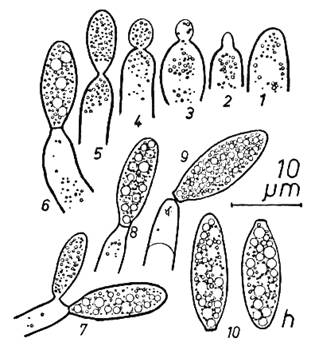
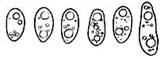
Conidial morphology. Conidia are comparatively small, bi- or
multiguttulate, and contain one or several nuclei (the nuclei are very difficult
to see). In contrast to the ascospores, the conidial wall does not stain in
CRB.
Germination
of conidia. Usually
no germination of the conidia of Sarcoscypha
was ever observed, though extensive studies on thousands of conidia were
undertaken (Harrington 1990: 429). However, in S. jurana the conidia
swell enormously on agar and eventually form a mycelium.
Blackish-brown
pigment of the branch surface.
In all species of Sarcoscypha
examined, a characteristic, more or less deep blackish-brown pigmentation of
the substrate surface was noted. In cultural studies with S. coccinea performed by P. Zinth (pers. comm.) on sterilized
natural substrate placed on a layer of sand, this dark pigment was formed at
the bottom and the top of the sand layer. Clearly this pigment is formed by the
mycelium of the Sarcoscypha, though
it does not occur on current agar media. The pigment forms a very thin layer
that undoubtedly embraces those areas of the branch being colonized by the
hyaline mycelium.