Asci
Ascus
base. The asci of Sarcoscypha and also other
Sarcoscyphaceae, to the present knowledge, consistently arise from simpe septa.
This is in contrast to the other families of the Pezizales, in which the asci
mostly arise from croziers.
Ascus
apex. See under “Ascus Apex
Morphology and Iodine Reaction in Ascomycetes” (in preparation).
Ascospores
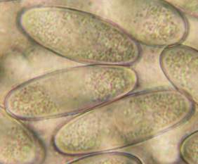
Shape. In S.
austriaca and S. jurana the
truncate, often indented (notched) poles have the shape of a saddle, so that a
turn of the spore for 90° along its longitudinal axis results in a
hemispherical to obtuse aspect of both poles. Therefore only about 50% of the
spores in a preparation show truncate poles (those seen in front view). This
fact is frequently mistaken as a variability in spore shape. If the spores are
dead, the degree of truncation or indentation is enhanced because of the
absence of an internal cell turgor. The function of this peculiar spore shape
is unknown; germination appears never to take place at the saddle but rather at
the edges (“shoulders”) or laterally.
|
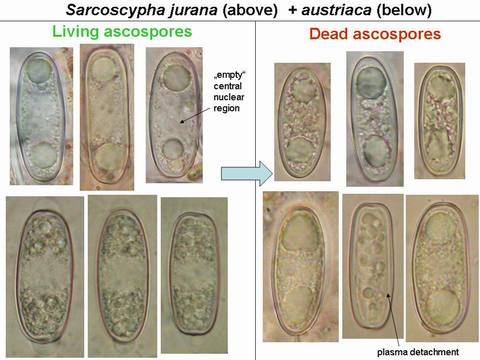
Mature ascospores of Sarcoscypha, living state |
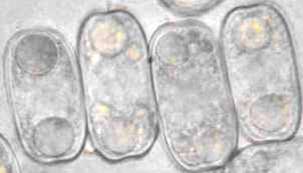
Mature ascospores of Sarcoscypha, living vs. dead state |
|
|
Sarcoscypha
austriaca (Salix, Tübingen, 31.3.2003, leg. H.O. Baral) |
Sarcoscypha
jurana (Tilia, Luxembourg, 16.3.2003, leg. G. Marson) |
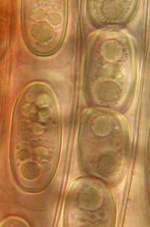
The strong difference in guttule pattern in the living spores between S. jurana and S. austriaca (left column) completely disappears in dead spores
due to coalescence of the oil drops, the spore interior thereby looking quite
variable (right column).
Therefore, herbarium material older than about 5-10 years is not easy to
identify. |
|
Sarcoscypha coccinea
(?macaronesica) (Quercus ilex, |
Sarcoscypha
coccinea (Ulmus, Montpellier, 22.2.2004, leg. G. Garcia) |
|
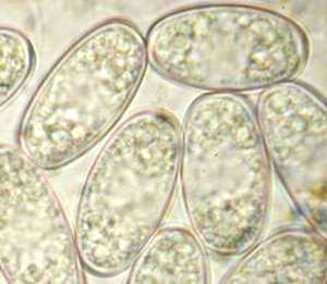
Contents. Characteristic
of the genus and perhaps the whole family is the presence of numerous
refractive minute oil drops (lipid bodies, LBs) among some larger LBs within the
living mature spores. A central area is free of LBs and contains 32 small
nuclei (?16 in S. occidentalis). Within Sarcoscypha only the
larger LBs are of taxonomic importance for species delimitation. While the
large and medium-sized LBs (characteristic of S. jurana and S. austriaca),
but also the minute LBs are rapidly seen in living spores, the relatively small
LBs of S. coccinea (see photo) and S. macaronesica are easily overlooked
among the minute ones. In dead spores the LBs are difficult to see because the
cytoplasma gets refractive when observed in a water mount. Mounting in KOH
makes the LBs reappear in full strength. However, coalescence of both larger
and smaller LBs takes place in many of the dead spores causing a very variable
guttule pattern which makes species recognition extremely difficult (see table
above). The specific guttule pattern is often already destroyed when the spore
powder got dry for only a few hours. On the other hand, some spores may still
show the unaltered guttule pattern even in very old herbarium material, helping
in recognizing the species.
|
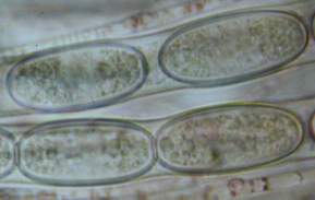
S.
jurana: spores inside living asci, with
mucilaginous envelope surronding complete spores. Envelope thick and swollen
in left ascus while compressed and high-refractive in right ascus. |
Mucilaginous envelope (sheath). In several of the species the mature ascospores
are surrounded by a delicate envelope. Within the living mature asci the
envelope is normally strongly compressed and of higher refractivity compared to
those spores within dead asci or being ejected. During immature stages,
however, an envelope is probably present in all taxa. The envelope is a very
delicate, evanescent structure which disappears in a watery environment some
period of time after ejection, and which is rather impossible to detect in
herbarium material. However, sheaths can well be seen in water mounts made from
spore prints several years after drying (Peric 2008). The envelope does not stain
in aqueous cresyl blue or Toluidin blue, while the spore wall surface stains
deep lilac in these reagents (only remnants of dead epiplasma around the
envelope stain violet).
The shape of the envelope
is rather constant within a species and serves as a valuable characteristic for
species delimitation. However, the envelope may be absent depending on the
geographical origin. S. austriaca usually has polar cap-like envelopes,
but sometimes no envelope at all. S. coccinea in Europe is completely
devoid of an envelope while in N-America an envelope around the whole spore
similar as in S. jurana is present
(Harrington 1990).