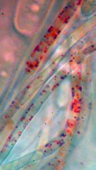
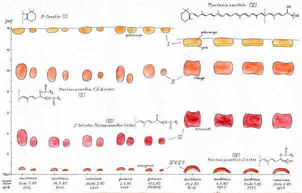
Paraphyses
and their pigment
The scarlet- to purple-red
hymenial colour of the disc is located in the paraphyses. It is caused by a
mixture of five different types of carotenoids (Arpin 1969, Baral 1983). This can
be demonstrated by thin layer chromatography. No difference in chemical
composition among the species of Sarcoscypha
examined could be detected. Carotenoids are lipidophilous and occur dissolved
in small lipid bodies (LBs) inside the paraphyses.
|
Carotenoids dissolved in LBs in
paraphyses of S. austriaca |
The five different types of carotenoids in
the three Central European species obtained by two procedures of thin layer
chromatography (IV and V are not recolved). |
|
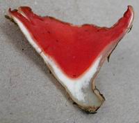
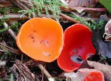
Sarcoscypha coccinea, red and yellow-orange form. France,
Chartres de Bretagne, Salix atrocinerea,
11.II.2004, A. Poncelet & J.P. Priou,
phot. JPP |
|
|
Sarcoscypha
jurana, white form, Côte d'Or, Tarsul, 23.XII.2008, P. Roger,
phot. A. Gardiennet |
|
 .
.
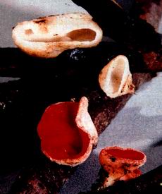
Albinism:
As an exception, groups of
apothecia were discovered with orange, yellow, and even white hymenia. Such
albinism can obviously be explained by the absence of one or more of these
carotenoids. The phenomenon is thought to be a genetically determined defect,
and appears quite rarely to occur. Apothecia with normal and reduced pigment
may sometimes grow side by side on the same branch (J.P. Priou, pers. comm., S. coccinea, see photo). On the polders
in Netherlands G. & Y. van Duuren showed me completely white apothecia of S. coccinea, and recently discovered
also such of S. austriaca. Finds of white
apothecia of S. jurana are known
Belgium and France (see figs. left). In herbarium specimens the hymenial colour
strongly fades with the years, thus making notes from the fresh state
necessary. A number of taxa have been described referring to this phenomenon
which occurs in all three species from
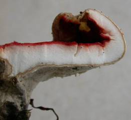
Medullary
and ectal excipulum, hairs
No difference in the composition
of the excipulum is known among the species of the genus. The medullary
excipulum forms the very thick “flesh” of the apothecia. It is a loosely interwoven,
non-gelatinized textura intricata of narrow hyphae. With its intercellular
space it serves as a water storage organ. The ectal excipulum is a compact
textura prismatica oriented at a very low angle to the surface. It gives rise
to long interwoven hairs forming a white felt on the exterior, especially on
the lower flanks and stipe. The curled to corkscrew-like shape of the hairs of S. austriaca was found to provide a
valuable character in separating this species from the other species of the
genus with more or less straight hairs (Baral 1984, Harrington 1990, Butterfill
& Spooner 1995, Pidlich-Aigner 1999: 17).
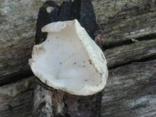
|
Median section through apothecium showing medullary
excipulum (above), ectal excipulum (centre), and hairs (below) |
||
|
Sarcoscypha austriaca
(Feldberg, Napf, Acer). Hairs flexuous to curled. |
Sarcoscypha
jurana (Lauterach, Wolfstal, Tilia). Hairs straight to wavy (usually not so curled). |
Sarcoscypha coccinea
(Karlsruhe, Hambrücken, Ulmus).
Hairs +/- straight. |
|
The medullary excipulum stores
extracellular water. Grey = intercellular space water-filled, white =
air-filled (S. jurana). |
State of complete loss of
extracellular water after c. 1 day exposure to a dry atmosphere. |